Spine Anatomy: Vertebrae, Discs and Nerves
The main regions
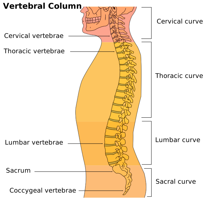
The spine is generally divided into the cervical (neck, 7 vertebrae), thoracic (upper/mid back, 12 vertebrae), lumbar (lower back, 5 vertebrae), plus the sacrum and coccyx at the base.
Discs and nerves
Between most vertebrae sit intervertebral discs — a tough outer layer around a softer, gel-like center — that cushion movement and absorb load. Spinal nerves branch out between vertebrae to the rest of the body, which is why problems at a specific spinal level can produce symptoms (numbness, pain) in a related area of the limbs.
Why this matters for common complaints
Understanding which spinal region and structure is involved helps explain why, for example, a lower lumbar disc issue can cause leg symptoms (as in sciatica), while a neck-level problem tends to affect the arms instead.
Key takeaways
- The spine has four main regions: cervical, thoracic, lumbar, plus the sacrum/coccyx.
- Discs cushion movement between vertebrae; nerves branching from the spine explain referred symptoms in the limbs.
- The spinal level involved often explains where symptoms show up in the body.
Sources
Related articles


This article is for general educational purposes only and is not medical advice. It does not replace diagnosis, treatment, or personalized recommendations from a licensed healthcare professional. Hands On Health is an independent blog and is not affiliated with any clinic.